
A groundbreaking cryo-electron microscopy image shows in extreme detail how a novel therapeutic molecule interacts with a protein, giving researchers insights into how it works in the human body. The molecule, ISRIB, was shown in a recent study by UCSF's Peter Walter, PhD, and Susanna Rosi, PhD, to restore memory failure in patients with traumatic brain injury. Image by the Adam Frost Lab
UC San Francisco researchers recently captured exquisite images of a protein caught in the act of binding to a novel therapeutic drug with enough resolution to model how the individual atoms of the protein and drug lined up.
Until recently, such a feat would have been considered impossible, but in the past five years, such breakthroughs have become almost commonplace here, part of a resolution revolution being led by UCSF researchers.
Thanks to their recent advances in cryo-electron microscopy (cryo-EM) – a technique whose inventors were honored with the 2017 Nobel Prize in Chemistry – these researchers have enabled rapid progress in the search for more precise and powerful therapies for a wide array of human diseases.
Proteins are tiny molecular machines that power everything our cells do – and they are also the primary targets for pharmaceutical drugs. Discovering how new proteins work could lead to lifesaving therapies for cancer or new painkillers with less danger of addiction.
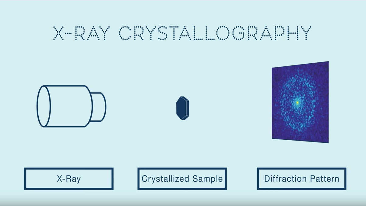
But to truly understand how proteins function, researchers must be able to visualize them at an atomic scale. Previously this required painstakingly growing crystals of a protein – a process which could take years to get right – then shooting X-rays through the crystals to calculate the protein’s atomic composition. Unfortunately, many of the most interesting proteins and complexes cannot be crystallized.
Cryo-EM has been around since the 1970s, but since 2013 technical advances pioneered in part by UCSF’s David Agard, PhD, and Yifan Cheng, PhD, have dramatically improved cryo-EM’s ability to resolve both the tiniest and the most complex of proteins in stunning detail, transforming the electron microscope from an old-fashioned back-of-the-lab workhorse to the newest scientific super-star almost overnight.