The Preclinical Therapeutics Core (PTC) offers a complete set of preclinical services and in vivo imaging devices for cancer investigators at UCSF and beyond.
Core Overview
The Preclinical Therapeutics Core (PTC):
- Conducts preclinical oncology trials for UCSF Helen Diller Family Comprehensive Cancer Center investigators and industry partners.
- Offers an extensive set of in vivo services covered under a comprehensive IACUC protocol, and provides training for common animal procedures and survival surgeries.
- Oversees and provides access and user training for multiple small animal imaging devices housed within the barrier facility.
- Manages and provides user training for various irradiator technologies, including precision irradiation.
- Imaging projects and irradiation experiments can be run entirely by Core personnel, if requested.
The Core utilizes two levels for animal hygiene, a standard specific pathogen free (spf) facility for immunocompetent mice, and a superclean facility with additional precautions to prevent infection of immunocompromised mice, such as those used to grow human tumors in vivo.
In experiments where the immune system is not involved, animals are housed in a regular room in the same building (HD525). In here, immunocompetent mice are used to conduct experiments with murine cancer cells.
The Core manages a room dedicated for imaging studies (HD527). Imaging instruments in this room are accessible to all members of the UCSF scientific community.
Workflow, Services, and Equipment
In vivo Services
The PTC offers an extensive set of in vivo services covered under a comprehensive IACUC protocol, and provides training for common animal procedures and survival surgeries.
- The PTC has expertise in high volume cell culture, and maintains a large cryorepository of commonly used tumor cell lines and tissues.
- Xenografts or allografts are generated via cell and/or tissue implantation; as well as orthotopic engraftment in different mouse strains depending on the required animal immune status. The cells and/or tissue sample can be implanted in different locations based on the desired animal model. In example, subcutaneous, intramammary fat pad, intracranial, intratibial, etc.
- The PTC has expertise in formulating experimental agents for delivery using a variety of commonly used methods (i.e. oral gavage, intra-peritoneal, intra-venous (tail vein or retroorbital injections), hydrodynamic, intra-tumoral, subcutaneous and osmotic mini-pumps).
- Mice are routinely monitored by body weight and physical observation following institutional welfare guidelines. Tumor measurement is preformed using digital calipers.
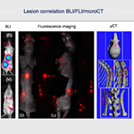
- In vivo imaging is also used to assess tumor burden, location, and dissemination. Available imaging technologies include μCT, ultrasound, and optical imaging.
- PTC performs collection of body fluids (including blood, thoracic, and peritoneal effusions and urine), tissues, and tumors as needed per experimental design. Standard tissue processing includes fixation for paraffin/OCT embedding, snap-freezing, and cryopreservation.

- The Core assists the scientific community in their PD and PK studies under request.
- For PK analysis, after single or multi-day delivery of experimental agent, plasma/serum is collected at multiple time points over a 24-72 hr period for assessment of drug persistency (note: analyte determination is run by user).
- For PD analysis, animals are typically treated with an experimental agent for 3-5 days before tissue/tumor collection for molecular target analysis or biodistribution.

- The Core engrafts fresh human tumor tissue derived from patient biopsy or surgery into immunodeficient mice in the clean area.
- After tumor growth, tissue is collected by surgical removal, dissected into multiple fragments, and reimplanted into a new batch of host mice. Other fragments are cryopreserved and banked for further work.
- The PTC generates new and established models of metastatic disease for cancer research. This is done by intravenous injection of cancer cells in most of the times, but intracardiac injection can be performed too, with ultrasound guidance. In most cases, molecular imaging methods allow evaluation of tumor location and overall tumor burden, including metastases.
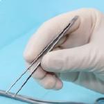
- The PTC in collaboration with LARC has developed a program of certified training for survival surgery procedures. If required, Core staff can preform the surgeries for the users in case emergency or lack of trained lab personnel.
- The PTC provides both 1:1 sessions and hands-on training to assist students and research fellows who need to learn surgical procedures.
Imaging Services
The PTC oversees and provides access and user training for multiple small animal imaging devices housed within the barrier facilities at Mission Bay (HD503 and HD527), Mount Zion (S0171) and Parnassus (PSB554) campuses. If necessary, PTC can run the imaging experiments by request.
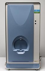
- The IVIS® Spectrum carries out high sensitivity and versatility optical-imaging studies. It is defined as a lab animal dedicated imager for light analysis and can analyze quantitatively bioluminescent (BLI) and fluorescent (FLI) signals.
- BLI studies require a previous genetic modification of the cells for introducing the luciferase gen. Once expressed, the injection of luciferin substrate triggers a specific intracellular reaction that generates photons of light that will be measured by the IVIS. The bioluminescence technique is ideal for making clear the expression of genes labeled with luciferase enzyme.
- FLI imaging is based on the tracking of specific fluorescent proteins (fluorophores). The IVIS possesses various fluorescent filters which cover most part of the light spectra, from yellow range to infrared dots. This provides great versatility to the research and enables countless biological or artificial structures to be marked and monitored.
- There is also the possibility of running ex vivo or in vitro studies, which provides optical imaging with an extensive range of application, from the first stages of experimental development to the final steps of the pre-clinical stage.
- This tool is optimal for application on oncology, microbiology, immunology or drug development.
- Easily and efficiently obtain calibrated quantitative data in your animal models of disease.
- Obtain functional and biological data to improve study designs and enhance decision making.
- Manage costs and capture time course data by avoiding sacrifice of animal models.
- Dual illumination capability enables topographic localization of both shallow and deep tumors in 3-D and reduces background interference.
- 20µM Resolution at 3.9cM field width
- In a similar way to IVIS Spectrum, IVIS Lumina offers the possibility for cell and proteins tracking with both BLI and FLI.
- This instrument is located in the clean area (HD503) giving support to the experiment conducted in immunodeficient animals.
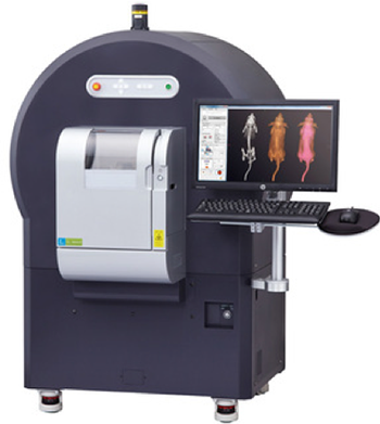
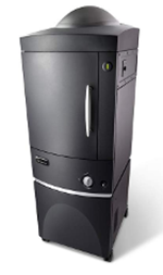
- A clinical device translated into lab animal’s size. The scanning bed and technical parameters are adapted to rodents and the system provides anatomical and functional information.
- Ideal for lung and bone studies, but also useful in soft tissue analysis using contrast agents.
- High resolution (2.3 uM voxel size) and High-speed scanning (scans as fast as 3.9 seconds)
- Low-dose imaging for longitudinal in vivo studies, avoiding the potential radiotherapeutic effect on the animals and allowing for multiple scans on the same animal (longitudinal experiments).
- Four Field of Views (FOVs) –18, 36, 72, and 86 mm.
- Two-phase retrospective respiratory and cardiac gating, ideal for imaging heart and lungs. Seamless co-registration of functional optical signals (from IVIS® Spectrum or FMT®) with microCT imaging data Multispecies imaging capabilities (Zebrafish/mouse/rat/guinea pig).
- The use of contrast agents increases the applicability of the equipment, including functional studies of the heart, analysis of the tissue perfusion or tumor vascularization.
- For getting access to this instrument, a specific radio-safety training is required. For more information, please contact the PTC.
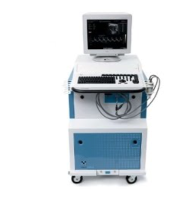
- Clinical technology adapted to lab animal. Ultrasonography is a completely harmless technique that enables real-time assessments of the structures. It is specially designed for cardiac and abdominal studies but can be used for any soft tissue or structure. Its speed of use and high versatility make an ideal tool for carrying out screenings in in vivo research.
- Non-invasive, non-radioactive, therapeutic regimes can be monitored in the same animal over time with resolution as low as 30 microns.
- Doppler modes allow for vascular anatomical identification (Power Doppler mode) and blood flow quantification (Pulsated wave Doppler).
- Possibility of tridimensional acquisitions and Volume analysis of structures.
- Micro vessels can be monitored using specifically designed contrast agents as well as targeted molecular biomarkers.
- Reduced applicability on brain or lung tissues.
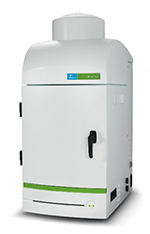
- A platform for tomographically quantitating in vivo optical imaging signals. FMT will efficiently provide quantitative, insightful data in your disease areas taking advantage of its 2 fluorescent channels.
- FMT preserves a linear relationship between activity in vivo and detector signal when imaging deep (non-surface) targets by reconstructing three-dimensional maps of fluorophores inside living animals.
- Using the FMT you can generate non-invasive, deep tissue quantitative data for pre-clinical research applications.
- Easily and efficiently obtain calibrated quantitative data in your animal models of disease
- Measure and monitor multiple biological processes simultaneously
- Obtain functional and biological data to improve study designs and enhance decision making
- Generate 3D, information-rich results
- Manage costs and capture time course data by avoiding sacrifice of animal models
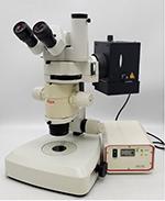
- Animal Surgery Stereo Microscope from Leica Microsystems. It provides a real-time high-resolution imaging combined with great depth of field for precision animal surgery or necropsy.
- With different fluorescent filters, including GFP and dsRed for green and red wavelengths fluorophores tracking.
Radiation Devices
The Core manages two different radiation devices located in the Helen Diller Family Comprehensive Cancer Center, inside the animal facility (HD537), which ensures a controlled environment for animal work.
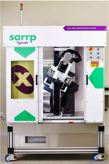
- A system specifically designed for lab animals, equivalent to the radiotherapy devices used in clinical oncology. It’s composed by a microCT imaging system for tissue density maps generation and a single beam irradiator for focal irradiation. Both parts combined create a singular and unique system in the cancer research.
- The SARRP can target micro beams (down to 0.5mm) of radiation achieving an accuracy of 200um for irradiated target.
- SARRP software allows user to image animal, contour the tumor/target and at-risk organs, evaluate dose, and deliver the desired treatment.
- With the multispecies platform, three animals can be imaged at the same time, which increases the throughput of the instrument and reduces the cost per animal.
- Because of its complexity, the use of the instrument SARRP is restricted to the Core staff. The PTC offers a complete service to the scientific community, including animal managing, image acquisition, and irradiation of the animals.

- A global X-ray based irradiator, user friendly and safe to use.
- From cells to small animals, the X-Rad320 delivers a precise radiation dose to specimens.
- With a large internal x-ray chamber, which permits to irradiate multiple cells-plates or mice at the same time.
- Full screen, real-time specimen viewing.
- For getting access to this instrument, a specific radio-safety training is required. More information can be obtained contacting the PTC.