

About LCA Microscopy
The LCA has several digital microscopes available to users including state-of-the-art laser-scanning and spinning disc confocal microscopes, high-throughput microscopy, and laser microdissection on the Mission Bay campus in the Hellen Diller Cancer Research Center and CVRB. Imaging consultations and analysis assistance is also available.
Instrument Operation/Training Rates:
Please refer to our iLab page for up to date hourly rates: LCA
For inquiries or operator and technical assistance, please contact Anna Celli at [email protected].
BC43 Spinning Disc Confocal
Benchtop multimodal microscope for widefield, brightfield, differential phase contrast, high-speed confocal, and optional super resolution. This microscope features four confocal and epifluorescence channels, environment control for live-cell time lapse imaging and high sensitivity for semi thick tissue imaging.
Instrument features:
- Configuration: Inverted
- Excitation: four laser lines (405nm, 488nm, 561nm, 638nm)
- Confocal module: Andor
- Emission: Blue channel: BP 435-455nm; Green channel: BP 517-541nm; Red channel: BP 580-610nm; Far Red channel: BP 671-745nmDetection: 4.1 MP (6.5 μm pixel, 16-bit) monochrome camera with up to 82% QE. 18.5 mm diagonal field of view.
- Objectives:
- 2X for sample navigation
- 10X Plan Apo LD Air Object 0.45NA 4.0WD
- 20x S Plan Fluor Objective 0.7NA 2.3 WD
- 40x Plan Fluor Oil Objective 1.3NA 0.24mm WD
- 60X Plan Apo LD Oil Object 1.42NA 0.15WD
- Operating system: Fusion
Sample table:
| Live samples | Semi thick samples | Thick tissue | Fixed slides | Culture plates |
| yes | yes | no | yes | yes |
Application table:
| Z-stack | Multichannel | Tiling and multiposition | Epifluorescence | Color camera (IHC) | Highthroughput /high content | Timelapse | Environment control |
| yes | yes | yes | yes | no | no | yes | yes |
Location: HD356 (inner room), Helen Diller Building, Mission Bay Campus
SOPs and required trainings:
- Training is required prior to first use
- Reservation through iLab is mandatory. Click here: iLab Organizer
Contact: For operator and technical assistance on this microscope, please contact Anna Celli, PhD at [email protected].
Rates: Please refer to our iLab page for up to date hourly rates: LCA
Zeiss Cell Observer Spinning Disc Confocal
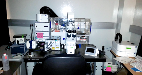
This spinning disk confocal microscope features full environmental control for long time-lapse multichannel live cell and tissue imaging, software controlled motorized stage and objective turret for large area tiling and multi position/well acquisition and z-stacking.
Instrument features:
- Configuration: Inverted
- Excitation: three laser lines (405nm, 488nm, 561nm), and an X-Cite 120Q mercury lamp
- Confocal module: Yocagawa CSU X1
- Emission: confocal arm - DAPI, GFP, mCherry; Epifluorescence arm- DAPI, GFP, mCherry, Cy5. Click here for system spectral configuration (Add link to FB database).
- Detection: Confocal arm- Evolve EMCCD; epifluorescence arm- AxioCam 506M
- Objectives: 2.5X Plan-Neo, 10X A Plan, 20X Plan-Apo, 40X LD Plan-Neo, 63X
- Operating system: ZEN blue 2012 (Carl Zeiss)
Sample table:
| Live samples | Semi thick samples | Thick tissue | Fixed slides | Culture plates |
| yes | yes | no | yes | yes |
Application table:
| Z-stack | Multichannel | Tiling and multiposition | Epifluorescence | Color camera (IHC) | Highthroughput /high content | Timelapse | Environment control |
| yes | yes | yes | yes | no | no | yes | yes |
Location: HD356 (inner room), Helen Diller Building, Mission Bay Campus
SOPs and required trainings:
- Training is required prior to first use
- Reservation through iLab is mandatory. Click here: iLab Organizer
Contact: For operator and technical assistance on this microscope, please contact Anna Celli, PhD at [email protected].
Rates: Please refer to our iLab page for up to date hourly rates: LCA
Zeiss Confocal LSM 780
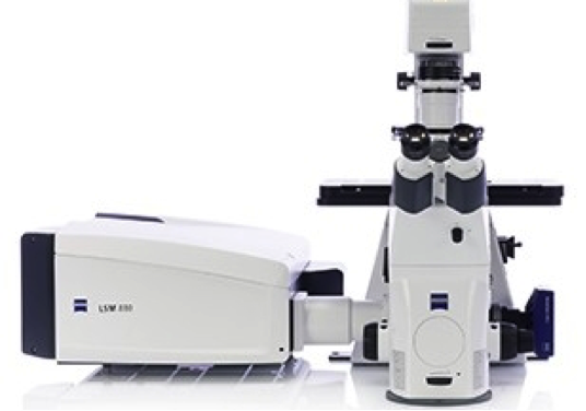 The Zeiss Confocal Laser-Scanning Microscope 780 is a scanning confocal and 2-photon microscope specifically suited for in vivo imaging of live animals and deep tissue imaging of live and fixed samples. Live cell imaging is available using a high NA 20X dipping lens. Detectors include two sensitive external GaAsP detectors for 2-photon imaging and Fluorescence Lifetime Imaging (FLIM), and three fully spectral internal detectors, including a 34 channel GaAsP array for detection of multicolor-labeled samples.
Location: CVRB Room 559, Cardiovascular Research Institute, Mission Bay Campus
SOPs and required trainings:
- Training is required prior to first use
- Reservation through iLab is mandatory. Click here: iLab
Contact: For operator and technical assistance on this microscope, please contact Anna Celli at [email protected]
Rates: Please refer to iLab for up to date rates LCA
Instrument features:
- Objectives: 5X, 10X LD, 20X dipping 2mm WD, 25X immersion, 63X water
- Light path: Upright
- Live Imaging Capabilities: Yes (no environmental control for cell cultures)
- Excitation (Lasers or filters): Laser lines: 405nm, 458nm, 488nm, 514nm, 633nm
- LED lamp for sample visualization: DAPI, GFP, TxRed
- Multi-photon Excitation: Mode-locked IR laser (690-1040nm) Chameleon Ultra II
- LED lamp for sample visualization (DAPI, GFP, TxRed)
- Confocal Type: Point-scanning
- Software Platform: ZEN
Sample table:
| Live samples | Semi thick samples | Thick tissue | Fixed slides | Culture plates |
| yes | yes | yes | yes | No, only 35mm-60mm dishes |
Application table:
| Z-stack | Multichannel | Tiling and multiposition | Epifluorescence | Color camera (IHC) | Highthroughput /high content | Timelapse | Environment control |
| yes | yes | yes | Only for sample localization | no | no | yes | Only temperature |
Agilent Cytation 5
The Agilent Cytation 5 is a versatile, automated imaging and multi-mode plate reader designed for high-content cellular analysis. Combining fluorescence and brightfield microscopy with multi-mode detection, it enables researchers to capture detailed cellular images and perform quantitative assays in the same instrument. With interchangeable objectives, automated focusing, and temperature control, it supports live-cell imaging and short-term studies. Its broad wavelength range accommodates diverse fluorophores, while integrated software streamlines acquisition, analysis, and data management.
Location: HD330-LCA bench space, Helen Diller Building, Mission Bay Campus
SOPs and required trainings:
- Training is required prior to first use
- Reservation through iLab is mandatory. Click here: iLab
Contact: For operator and technical assistance on this microscope, please contact Anna Celli at [email protected]
Rates: Please refer to iLab for up to date rates LCA
Instrument features:
- Objectives: 1.25X, 4X, 10X LD, 20X , 40X, 60X (all dry)
- Light path: Inverted
- Channels: Several interchangeable cubes are available. Standard configuration: DAPI, EGFP, mCherry, autofocus. Additional cubes: Cy3, Cy5, Cy7
- Live Imaging Capabilities: Yes (no environmental control for cell cultures)
- Software Platform: Agilent Gen5
Sample table:
| Live samples | Semi thick samples | Thick tissue | Fixed slides | Culture plates |
| yes | no | no | yes | yes |
Application table:
| Z-stack | Multichannel | Tiling and multiposition | Epifluorescence | Color camera (IHC) | Highthroughput /high content | Timelapse | Environment control |
| yes | yes | yes | yes | yes | yes | yes | Only temperature |
Zeiss Observer
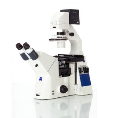 The Zeiss Observer is a basic tissue culture microscope for general-use. It is for use in all areas of light microscopy, including transmitted and incident light microscopy, fluorescent microscopy, darkfield, phase, and polarization contrast.
Location: HD356, Helen Diller Building, Mission Bay Campus
Contact: For operator and technical assistance on this microscope, please contact Anna Celli at [email protected]
Rates: Please refer to iLab for up to date rates LCA
Instrument features:
- Objectives: 10X Plan Neo, 20X LD A-Plan, 25X Plan-Neo, 40X LD A-Plan
- Light path: Inverted
- Excitation: X-Cite mini Lamp
- Channels: Dapi, GFP, RFP
- Software Platform: ZEN
Zeiss AxioImager 2
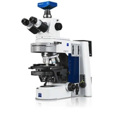
The Zeiss AxioImager 2 with AxioVision Software is a general-use, epiflurescent microscope for use in all areas of light microscopy, including transmitted and incident light microscopy, fluorescent microscopy, darkfield, phase, and polarization contrast.
Location: S171, Cancer Research Building, Mt Zion
SOPs and required trainings:
- Reservation through iLab is mandatory. Click here: iLab
Contact: For operator and technical assistance on this microscope, please contact Anna Celli at [email protected]
Rates: Please refer to iLab for up to date rates LCA
Instrument features:
- Objectives: 2.5X Plan-Neo, 10X Plan-Neo, 20X Plan-Apo, 40X Plan-Apo oil, 63X Plan-Apo oil
- Light path: Upright
- Excitation (lasers or filters): Lamp Dapi, GFP, Rhodamine, mCherry, Cy5
- Software Platform: AxioVision
Zeiss AxioImager M1
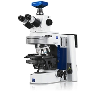
The Zeiss AxioImager M1 with AxioVision Software is a general-use, epiflurescent microscope for use in all areas of light microscopy, including transmitted and incident light microscopy, fluorescent microscopy, darkfield, phase, and polarization contrast.
Location: HD355, Helen Diller Building, Mission Bay Campus
Contact: For operator and technical assistance on this microscope, please contact Anna Celli at [email protected]
Rates: Please refer to iLab for up to date rates LCA
Instrument features:
- Objectives: 1.25X Plan-Neo, 5X Plan-Neo, 10X Plan-Neo, 20X Plan-Apo, 40X Plan-Neo, 63X Plan-Apo oil
- Light path: Upright
- Excitation (lasers or filters): Lamp Dapi, GFP, DsRded, mPlum, Cy5
- Software Platform: ZEN
Sample table:
| Live samples | Semi thick samples | Thick tissue | Fixed slides | Culture plates |
| no | no | no | yes | no |
Application table:
| Z-stack | Multichannel | Tiling and multiposition | Epifluorescence | Color camera (IHC) | Highthroughput /high content | Timelapse | Environment control |
| no | yes | yes | yes | yes | no | no | no |
Keyence BZ-x800
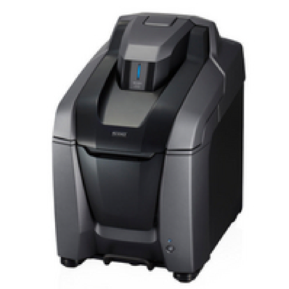
The Keyence BZ-X800 is a fully motorized, inverted all-in-one 4-channels fluorescence microscope combining fluorescence, brightfield, and phase-contrast imaging. It supports slides, dishes, well-plates, and flasks, with a motorized stage, lenses, and filter switching for seamless operation. Features include auto-focus, image stitching, time-lapse, large-area scanning.
Location: HD256, Helen Diller Building, Mission Bay Campus
SOPs and required trainings:
- Training is required prior to first use
- Reservation through iLab is mandatory. Click here: iLab
Contact: For operator and technical assistance on this microscope, please contact Anna Celli at [email protected]
Rates: Please refer to iLab for up to date rates LCA
Instrument features:
- Objectives: 4X, 10X LD, 20X, 40X
- Light path: Inverted
- Channels: DAPI, EGFP, mCherry, Cy5
- Live Imaging Capabilities: Yes (no environmental control for cell cultures)
- Software Platform: Keyance
Sample table:
| Live samples | Semi thick samples | Thick tissue | Fixed slides | Culture plates |
| yes | no | no | yes | yes |
Application table:
| Z-stack | Multichannel | Tiling and multiposition | Epifluorescence | Color camera (IHC) | Highthroughput /high content | Timelapse | Environment control |
| yes | yes | yes | yes | yes | no | no | no |
Nikon Eclipse
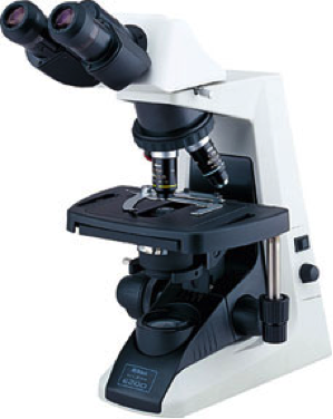 The Nikon Eclipse is a basic upright dual viewing teaching microscope.
Location: HD355, Helen Diller Building, Mission Bay Campus
Contact: For operator and technical assistance on this microscope, please contact Anna Celli at [email protected]
Rates: Please refer to iLab for up to date rates LCA
Instrument features:
- Objectives: 4X plan, 10X Plan, 20X Plan, 40X Plan
- Light path: Upright
- Excitation (lasers or filters): Bright field.
Zeiss PALM Laser Microdissection
The Zeiss PALM Laser Microdissection system offers contact free manipulation and laser driven high precision tissue microdissection and capture. This is a tool for isolation of single cells or cell groups for downstream genomics, transcriptomics, and/or proteomics analysis and applications.
Location: CVRB room 559, Cardiovascular Research Building, Mission Bay Campus
SOPs and required trainings:
- Training is required prior to first use
- Reservation through iLab is mandatory. Click here: iLab
Contact: For operator and technical assistance on this microscope, please contact Anna Celli at [email protected]
Rates: Please refer to iLab for up to date rates LCA
Instrument features:
- Objectives: 5X Fluar, 10X Fluar, 20X LD Plan-Neo, 40X LD Plan-Neo, 63X Plan-Neo
- Light path: Inverted
- Excitation (lasers or filters): Lamp Dapi, GFP, Rhoda mine, Cy5
- Other features: Touch-cap collection
- Software Platform: Palm Robot
Sample table:
| Live samples | Semi thick samples | Thick tissue | Fixed slides | Culture plates |
| no | no | no | yes | no |
Application: Laser-activated microdissection and capture of user-defined regions on fixed and frozen sections, or fixed cells for down stream proteomics, genomic, or transcriptomic analysis.
Data Analysis workstation
The LCA offers a workstation for post acquisition microscopy and flow cytometry data analysis.
Location: HD561, Helen Diller Building, Mission Bay Campus
SOPs and required trainings: Reservation through iLab is mandatory. Click here to reserve the workstation through iLab: iLab Organizer
Contact: For operator and technical assistance on this microscope, please contact Anna Celli at [email protected], Sarah Elmes at [email protected], or Danielle Peterson at [email protected]
Analysis Softwares: Imaris (3D image segmentation and analysis), FlowJo (Flow cytometry data analysis), ImageJ, Cell Profiler, Zen lite (free image browsing of .czi files), Gen5 lite.